human ear, organ of hearing and equilibrium that detects and analyzes noises by transduction (or the conversion of sound waves into electrochemical impulses) and maintains the sense of balance (equilibrium). Hence called as the Stato acoustic organ of the body.
Anatomically Human ear mainly consists of 3 parts
- the outer part
- middle part
- Inner part
The Outer Part (Outer Ear)
- The outer most part of mammal is represented by the auricle. In humans the auricle is an almost rudimentary, usually immobile shell that lies close to the side of the head.
- In human Auricle is represented by ear pinna. It consists of a thin plate of yellow elastic cartilage covered by closely adherent skin. The cartilage is molded into clearly defined hollows, ridges, and furrows that form an irregular, shallow funnel.
- The ear pinna at its deepest part leads directly to the external auditory canal, or acoustic meatus, is called the Concho.
- The auditory canal carries sound to the eardrum, and its lining produces ear wax to keep the eardrum and canal from drying out and to trap dirt before it gets to the eardrum.
- The ear drum or Tympanic Membrane is the inner most part of the outer ear.
- When sound waves vibrate the eardrum, sound energy is transferred to the middle ear.
Middle Part (Middle Ear)
- Anatomically Middle ear is the 2nd part of the auditory system.
- The middle ear is a small, air-filled pocket bounded by the eardrum on one side and the oval window of the inner ear on the other.
- This pocket is connected to the common mouth and nasal cavity, or pharynx, by the Eustachian tube.
- The Eustachian tube allows air pressure to equalize between the outside of the eardrum (surrounding atmosphere) and the inside of the eardrum (the middle ear).
- The middle ear has three small bones in the body,
- the malleus – Hammer Shaped
- incus – Anvil shaped
- stapes – Stirrup Shaped
- The three bones form a chain of levers connected by joints. The malleus is attached to the eardrum by ligaments, as is the stapes to the oval window.
- this series of membranes and bones forms a very effective pathway that carries vibrations from the eardrum to the inner ear.
- The stapes, Vibrates the membranous oval window when the eardrum and the three bones are vibrated by sound waves.
- the oval window is a closed membrane, which acts as the entrance to the inner ear for sound energy.
Inner Part (Inner Ear)
The internal ear composed of these following parts
- oval window –it connects the middle ear with the inner ear
- semicircular ducts – it is filled with fluid; attached to cochlea and nerves; send information on balance and head position to the brain
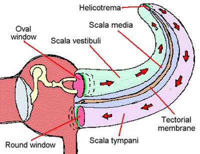
- cochlea – spiral-shaped organ of hearing; transforms sound into signals that get sent to the brain
- auditory tube – drains fluid from the middle ear into the throat behind the nose.
- Internal ear may be divided into 2 parts, namely Bony Labyrinth & Membranous Labyrinth.
- The bony labyrinth, or osseous labyrinth, is the network of passages with bony walls lined with periosteum.
- The membranous labyrinth runs inside of the bony labyrinth. There is a layer of perilymph fluid between them. The three parts of the bony labyrinth are the vestibule of the ear, the semicircular canals, and the cochlea.
- The cochlear canals contain two types of fluid: perilymph and endolymph.
- Perilymph and Endolymph devices the Cochlea into three distinct parts.
- scala tympani
- Scala vestibuli
- Scala media


